CLINICAL APPLICATIONS
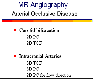
Arterial Occlusive Disease
A major application for MRA is screening the carotid arteries for arteriosclerotic disease.
![]() Stroke
is the third most common cause of death in the United States. A common source of TIA and stroke
is atheromatous disease at the carotid bifurcation in the neck. Since the NASCET (North American
Symptomatic Carotid Endarterectomy Trial) study
Stroke
is the third most common cause of death in the United States. A common source of TIA and stroke
is atheromatous disease at the carotid bifurcation in the neck. Since the NASCET (North American
Symptomatic Carotid Endarterectomy Trial) study
![]() has proved the efficacy of carotid endarterectomy for stenoses ≥70%, accurate
assessment of the carotid artery is of paramount importance.
has proved the efficacy of carotid endarterectomy for stenoses ≥70%, accurate
assessment of the carotid artery is of paramount importance.
Gd-enhanced MRA has replaced TOF and PC methods for imaging the neck vessels. It is faster
and more robust than the non-contrast techniques. The ability to visualize the arteries from their
origins at the aortic arch all the way to the circle of Willis makes it possible to do a complete vascular
assessment. Gd-enhanced MRA is far superior for imaging high-grade stenoses and for visualizing
slow distal flow. Signal loss from flow stasis or swirling within the carotid bulb is not a problem with
this technique. Since no superior SAT pulses are needed to suppress venous structures, no saturation
occurs in vascular loops.
![]()
Another application of MRA is in cases of suspected dissection. Relatively minor trauma is
sufficient to cause a dissection, or it can be spontaneous. The MRA may demonstrate complete
occlusion or only narrowing of the arterial lumen. Spin-echo images should also be obtained because
they are very sensitive for detecting the intramural hemorrhage. The typical appearance of an oval-shaped hyperintensity with an eccentrically placed flow void may be more convincing for a dissection
than the MRA. The MRA is very useful for following a dissection to look for recanalization of a
complete occlusion or resolution of the vascular compromise caused by the intramural thrombus.
![]()
MRA can also evaluate the major intracranial arteries about the circle of Willis.
![]() Most protocols
use a 3D TOF sequence. Using MOTSA (multiple overlapping thin slabs) with 3D TOF techniques
reduces saturation effects and allows visualization of more distal arteries. Intracranial MRA is a very
acceptable method for imaging the vertebrobasilar system, which is inaccessible to ultrasound and has
no effective surgical therapy.
Most protocols
use a 3D TOF sequence. Using MOTSA (multiple overlapping thin slabs) with 3D TOF techniques
reduces saturation effects and allows visualization of more distal arteries. Intracranial MRA is a very
acceptable method for imaging the vertebrobasilar system, which is inaccessible to ultrasound and has
no effective surgical therapy.
![]() The phase images of 2D PC MRA can be used to determine direction
of collateral flow about the circle of Willis.
The phase images of 2D PC MRA can be used to determine direction
of collateral flow about the circle of Willis.
When reading MR angiograms, it is important to remember that non-contrast TOF techniques place an rf saturation (SAT) pulse above the imaging plane to eliminate signal from venous structures flowing inferiorly. If any arteries happen to flow from superior to inferior, they will also be masked. For example, retrograde flow in a vertebral artery secondary to a subclavian steal phenomenon will not be visualized. Similarly, retrograde filling of a carotid artery or the basilar artery in the presence of severe proximal disease may not be imaged and lead to misdiagnosis of occlusion. A vascular loop can also result in loss of signal. 2D TOF is more susceptible to these artifacts because the SAT band is placed about 5 mm above each successive image slice. Misdiagnosis of occlusion can be avoided in these situations by repeating the TOF sequence with no SAT band or by doing a Gd-enhanced MRA or a PC study.
{To return to cases, use the "Back " button on the Toolbar}