ABSCESS
Bacterial
Brain abscesses may be related to infections of the paranasal sinuses, mastoids, middle ears as well as hematogenous seeding, but in 20% of cases a source is not discovered. Very rarely an abscess is secondary to meningitis. In children, more than 60% of cerebral abscesses are associated with congenital heart disease and right to left shunts. Presenting symptoms of a cerebral abscess include headache, drowsiness, confusion, seizures and focal neurologic deficits. Fever and leukocytosis are common during the invasive phase of a cerebral abscess but may resolve as the abscess becomes encapsulated. Organisms most frequently cultured from brain abscesses in otherwise immunocompetent individuals are staphylococcus and streptococcus.
When the brain is inoculated with a pathogen, a local cerebritis develops. Pathologically, an area of cerebritis consists of vascular congestion, petechial hemorrhage and brain edema. The infection goes through a stage of cerebral softening, followed by liquefaction and central cavitation. With time, the central necrotic areas become confluent and are encapsulated after one to two weeks. Edema, a prominent feature of cerebral abscess, may actually subside after the capsule forms.
In the cerebritis stage, MR reveals high signal intensity on T2-weighted images, both
centrally from inflammation and peripherally from edema. Areas of low signal are variably
imaged on T1-weighted scans. As the progression to abscess ensues there is further
prolongation of T1 and T2 centrally. The capsule becomes highlighted as a relatively
isointense structure containing and surrounded by low signal on T1- weighted images, and
high signal on T2-weighted images. Mottled areas of enhancement are seen with gadolinium-enhanced MR during the cerebritis stage, with an enhancing rim developing as the abscess
matures. The enhancing rim may appear late in the cerebritis stage, prior to actual central
necrosis. In some instances, the central area of necrosis has also enhanced on delayed scans,
but not as commonly as is seen in necrotic tumors.
![]() ,
,
![]()
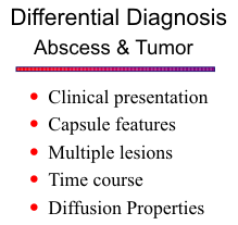
Advanced neuroimaging techniques, such as MR spectroscopy (MRS) and
diffusion-weighted imaging (DWI) , have markedly improved the specificity of MRI for
distinguishing bacteria abscess from other infections and from cystic and necrotic tumors.
MRS reveals metabolites of bacterial origin, including acetate, lactate, succinate, cytosolic
acid, and amino acids (alanine, valine, leucine). The spectral pattern of cystic or necrotic brain
tumors is quite different and normally contains elevated choline and decreased
N-acetyl-aspartate (NAA), with variable amounts of lactate and lipids. DWI shows restricted
diffusion and high signal intensity in bacterial abscesses. The presence of pus within the
abscess cavity, which consists of numerous
leukocytes and proteinaceous fluid with high
viscosity, accounts for the restricted diffusion and
high signal intensity on DWI and low ADC values. In
contrast, the cystic or necrotic portions of brain
tumors typically are less cellular and have less viscous
fluid consistency. As a result, tumors show low
signal intensity on DWI and higher ADC values.
![]()
{To return to cases, use the "Back " button on the Toolbar}