Lumbar Spine
Congenital spinal stenosis is often asymptomatic until middle age, when secondary degenerative changes develop. The acquired type is a disease of primarily adult men with moderate to severe degenerative spine disease. The syndrome of neurogenic or spinal claudication includes bilateral lower extremity pain, numbness, and weakness that is poorly localized and usually associated with low back pain. The symptoms are worse with standing or walking and relieved when the patient lies down.
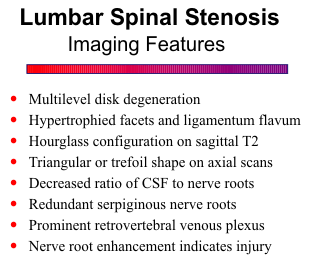
Spinal stenosis is graphically displayed in
the sagittal plane by gradient-echo or T2-
weighted pulse sequences. The hyperintense
thecal sac is effaced anteriorly by the bulging
disk and posteriorly by the ligamentum
flavum, resulting in an hourglass
configuration. Acquired spinal stenosis is
usually associated with moderate to severe
multilevel disk degeneration, consisting of
loss of normal signal, disk space narrowing,
and intradiskal calcification or air. The
calcification and air can be difficult to discern within severely desiccated disks. On axial views the
constricted canal often has a triangular or trefoil shape due to encroachment on the posterolateral
aspects of the canal by hypertrophied facets.
![]()

Since compression of the nerve roots within the thecal sac causes the symptoms, assessment of
the ratio of CSF to nerve roots is important to make the diagnosis of spinal stenosis. With
progressive stenosis, the amount of CSF progressively diminishes and the nerve roots become
crowded together. Constriction at the level of stenosis prevents the normal superior and inferior
movement of the nerve roots with flexion and extension, resulting in a redundant serpiginous root
pattern above and below the stenosis. Nerve root enhancement may also be seen, due to either
breakdown of the blood-nerve barrier from mechanical injury, inflammatory response, and Wallerian
degeneration/regeneration of axons, or engorgement of intrathecal veins and perineural vascular
plexus. As a result of epidural compression, prominent enhancement of retrovertebral venous plexus
is common.
![]()
{To return to cases, use the "Back " button on the Toolbar}