INFECTIOUS AND INFLAMMATORY DISORDERS
John R. Hesselink, MD, FACR
Inflammatory diseases of the brain include abscess, meningitis, encephalitis and
vasculitis. The brain is protected from invading infectious agents by the calvarium, dura and
blood- brain barrier. Moreover, the cerebral tissue itself is relatively resistant to infection.
Most pyogenic infections are hematogenous and related to septicemia and endocarditis.
Direct extension from an infected paranasal sinus or middle ear/mastoid is less common than
in the pre-antibiotic era. Fungal infections are less common than bacterial infections, but are
taking on more importance in AIDS patients and those immunocompromised by way of
chemotherapy, neoplasia, or immunosuppressive therapy for organ transplantation. The most
important viral infections of the central nervous system from an imaging point of view are
aseptic meningitis, encephalitis, and progressive multifocal leukoencephalopathy (PML).
Herpes simplex is responsible for a fulminant viral encephalitis, and both the human
immunodeficiency virus (HIV) and cytomegalovirus (CMV) produce a white matter
encephalitis associated with the AIDS epidemic.
![]() ,
,
![]()
ABSCESS
Bacterial
Brain abscesses may be related to infections of the paranasal sinuses, mastoids, middle ears as well as hematogenous seeding, but in 20% of cases a source is not discovered. Very rarely an abscess is secondary to meningitis. In children, more than 60% of cerebral abscesses are associated with congenital heart disease and right to left shunts. Presenting symptoms of a cerebral abscess include headache, drowsiness, confusion, seizures and focal neurologic deficits. Fever and leukocytosis are common during the invasive phase of a cerebral abscess but may resolve as the abscess becomes encapsulated. Organisms most frequently cultured from brain abscesses in otherwise immunocompetent individuals are staphylococcus and streptococcus.
When the brain is inoculated with a pathogen, a local cerebritis develops. Pathologically, an area of cerebritis consists of vascular congestion, petechial hemorrhage and brain edema. The infection goes through a stage of cerebral softening, followed by liquefaction and central cavitation. With time, the central necrotic areas become confluent and are encapsulated after one to two weeks. Edema, a prominent feature of cerebral abscess, may actually subside after the capsule forms.
In the cerebritis stage, MR reveals high signal intensity on T2-weighted images, both
centrally from inflammation and peripherally from edema. Areas of low signal are variably
imaged on T1-weighted scans. As the progression to abscess ensues there is further
prolongation of T1 and T2 centrally. The capsule becomes highlighted as a relatively
isointense structure containing and surrounded by low signal on T1- weighted images, and
high signal on T2-weighted images. Mottled areas of enhancement are seen with gadolinium-enhanced MR during the cerebritis stage, with an enhancing rim developing as the abscess
matures. The enhancing rim may appear late in the cerebritis stage, prior to actual central
necrosis. In some instances, the central area of necrosis has also enhanced on delayed scans,
but not as commonly as is seen in necrotic tumors.
![]() ,
,
![]()
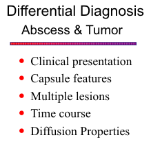
Advanced neuroimaging techniques, such as MR spectroscopy (MRS) and
diffusion-weighted imaging (DWI) , have markedly improved the specificity of MRI for
distinguishing bacteria abscess from other infections and from cystic and necrotic tumors.
MRS reveals metabolites of bacterial origin, including acetate, lactate, succinate, cytosolic
acid, and amino acids (alanine, valine, leucine). The spectral pattern of cystic or necrotic brain
tumors is quite different and normally contains elevated choline and decreased
N-acetyl-aspartate (NAA), with variable amounts of lactate and lipids. DWI shows restricted
diffusion and high signal intensity in bacterial abscesses. The presence of pus within the
abscess cavity, which consists of numerous
leukocytes and proteinaceous fluid with high
viscosity, accounts for the restricted diffusion and
high signal intensity on DWI and low ADC values. In
contrast, the cystic or necrotic portions of brain
tumors typically are less cellular and have less viscous
fluid consistency. As a result, tumors show low
signal intensity on DWI and higher ADC values.
![]()
Fungal
Fungal organisms can start as a meningitis or cerebral abscess, or can invade directly from an extracranial compartment. As mentioned above, fungal infections are primarily found in immuno compromised hosts. In immunocompetent patients, fungal abscesses tend to evolve more slowly than bacterial abscesses, but that is not the case in patients with deficient immunity. In general, there are no specific MR imaging features to distinguish the infecting agent.
Coccidioidomycosis is endemic to the central valley regions of California and desert
areas of the southwestern United States. Infection occurs by inhalation of dust from soil
usually heavily infected with arthrospores. Primary coccidioidomycosis, a pulmonary
infection, is followed by dissemination in only about 0.2% of immunocompetent patients.
Central nervous system involvement most often represents a meningitis, but cerebral abscess
and granuloma formation can also occur.
![]()
Mucormycosis is seen most often in patients with poorly controlled diabetes. It starts as a necrotizing vasculitis of the nose and sinuses, and spreads by direct invasion of adjacent facial compartments. Extension into the intracranial cavity occurs through the cribriform plate, superior orbital fissure, and basal foramina or indirectly via involvement of vascular structures. Once within the intracranial cavity, it produces a purulent meningitis, cerebral infarction from arterial occlusion, and acute cerebritis due to direct invasion of the olfactory tracts and inferior frontal and temporal lobes. Cranial nerve involvement and cavernous sinus thrombosis are common. The MR findings reflect the observed pathologic changes. Regions of meningeal and cerebral inflammation are hyperintense on T2 and proton density weighted MR images. Infarction and edema account for additional high signal parenchymal abnormalities. Gadolinium-enhanced T1-weighted scans show enhancement of the basal meningeal inflammation, as well as the adjacent cerebral involvement. Coronal scans are especially helpful to display the relationships of the meningeal process to the brain and adjacent extracranial compartments.
Aspergillosis is an aggressive opportunistic fungal infection. The organism gains entrance with inhalation of infected grains or dusts and results in primarily a pulmonary infection. Pathologic changes include a combination of suppuration and granulomas. Dissemination to the CNS may start as a basal meningitis, but the organism readily invades vascular structures and extends into the brain parenchyma.
Cysticercosis
Neurocysticercosis is the most frequently encountered parasitic infestation of the CNS. Originally endemic in underdeveloped countries, predominantly Latin America, Africa, Asia and some portions of eastern Europe, it is becoming increasingly frequent in North America in immigrant populations. Humans become accidental hosts for the larval stage of Taenia Solium, the pork tapeworm, by ingesting contaminated material. The eggs hatch in the stomach and larvae burrow through the gut wall and become distributed by the circulatory system. There is a predilection for involvement of the brain. Patients most often present with seizures, elevated intracranial pressure, focal neurologic abnormalities and altered mental status. Asymptomatic infections are common.
Four forms of neurocysticercosis are described: meningeal, parenchymal, ventricular
and mixed. In all locations, death of the larva provokes a more intense inflammatory
response, and in the case of an intraventricular lesion may lead to ependymitis. Parenchymal
lesions consist of small cysts, large cysts and calcified lesions. Small (approximately 1.5 cm.
in diameter) cysts may have a central area of relatively shorter T1 (isointense or hyperintense
to cortex) and are uniformly hyperintense on T2-weighted images. Large (4-7 cm) cysts are
usually multiloculated, adjacent to the subarachnoid space and may contain a mural nodule.
The presence of a mural nodule or a T2-hypointense rim in encapsulated lesions may correlate
with larval death. Visualization of calcified lesions has been variable with MR; overall there
is an advantage for CT in this regard. Sometimes, calcified lesions are surrounded by edema,
making them more conspicuous on MR. Basal cistern lesions can be difficult to identify but
have been visualized as areas of intermediate signal intensity on T1-weighted images.
Intraventricular cysticercosis results in deformable and mobile cysts that may cause
intermittent hydrocephalus.
![]()
MENINGITIS
Bacterial
Bacterial meningitis is an infection of the pia and arachnoid and adjacent cerebrospinal fluid. The outer arachnoid serves as a barrier to the spread of infection, but involvement of the subdural space can occur, resulting in a subdural empyema. This complication is more common in children than adults. The most common organisms involved are Hemophilus influenza, Neisseria meningitides (Meningococcus) and Streptococcus pneumoniae. Patients present with fever, headache, seizures, altered consciousness and neck stiffness. The overall mortality rate ranges from 5 to 15% for H. influenza and meningococcal meningitis to as high as 30% with streptococcal meningitis. In addition, persistent neurologic deficits are found in 10% of children after H. influenza meningitis and in 30% of patients with streptococcal meningitis.
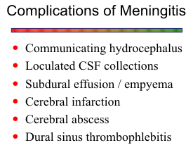
The ability of nonenhanced MR to image meningitis is extremely limited, and the
majority of cases are normal or have mild hydrocephalus. In severe cases, the basal cisterns
may be completely obliterated, with high signal intensity replacing the normal CSF signal on
proton density images. Intermediate signal intensity may be seen in the basal cisterns on T1-weighted images in these cases. Meningeal enhancement often is not present, unless a chronic
infection develops. Infection within the ventricles, either from direct extension from a shunt
or abscess or progression of meningitis, may
lead to ependymitis, resulting in
hyperintensity outlining the ventricles on T2-
weighted images and enhancement of the
ependyma on T1-weighted images with
gadolinium. Subdural empyemas are better
seen with MR than with CT, and the signal
characteristics of the exudate in subdural
empyema (higher signal than CSF) helps to
differentiate it from benign extra-axial
collections.
![]()
Tuberculosis
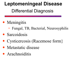
Tuberculous meningitis remains an important disease, becoming more common as an infectious agent in AIDS patients. As a rule, the evolution is less rapid than in pyogenic infections. Vasculitis and cerebral infarction, caused by inflammatory changes in the basal cisterns, are more prevalent. The MR features of tuberculous meningitis are similar to the bacterial agents, but the chronic inflammation induces thick granulation tissue that produces a more striking enhancement pattern. Actual intracranial tuberculomas are rare in the United States. Mature tuberculomas are T2 hypointense. Central necrosis in some lesions results in a T2 bright core with a low signal intensity rim.
Sarcoidosis
Sarcoidosis is a granulomatous disease of unknown etiology. In approximately 5% of cases, the CNS is involved as a granulomatous infiltration of the meninges and underlying parenchyma, most notably at the base of the brain. It may also affect cranial or peripheral nerves as isolated disease. Cranial nerve palsies, chronic meningitis and hypothalamic- pituitary dysfunction are frequent manifestations.
MR is well suited to imaging the
focal pituitary/hypothalamic lesions and white matter lesions that have been noted in these
patients. The basal cisterns may enhance in patients with meningeal sarcoidosis, but a nodular
pattern usually distinguishes it from the infectious varieties. A particularly interesting form
of meningeal sarcoid results in thick meningeal plaques, often over the convexities. These
may mimic meningiomas in that they remain isointense or hypointense relative to cortex on
T2- as well as T1-weighted images.
![]() ,
,
![]()
ENCEPHALITIS
Encephalitis refers to a diffuse parenchymal inflammation of the brain. Acute encephalitis of the non-herpetic type presents with signs and symptoms similar to meningitis but with the added features of any combination of convulsions, delirium, altered consciousness, aphasia, hemiparesis, ataxia, ocular palsies and facial weakness. The major causative agents are arthropod-borne arboviruses (Eastern and Western equine encephalitis, St. Louis encephalitis, California virus encephalitis). Eastern equine encephalitis is the most serious but fortunately also the least frequent of the arbovirus infections. The enteroviruses, such as coxsackie-virus and echoviruses, can produce a meningoencephalitis, but a more benign aseptic meningitis is more common with these organisms. MR reveals hyperintensity on T2-weighted scans within the cortical areas of involvement, associated with subcortical edema and mass effect.
Herpes Simplex
Herpes simplex is the commonest and gravest form of acute encephalitis with a 30-70% fatality rate and an equally high morbidity rate. It is almost always caused by Type 1
virus except in neonates where Type 2 predominates. Symptoms may reflect the propensity
to involve the inferomedial frontal and temporal lobes- hallucinations, seizures, personality
changes and aphasia. MR has demonstrated positive findings in viral encephalitis as soon as
2 days after symptoms, more quickly and definitively than CT. Early involvement of the
limbic system and temporal lobes is characteristic of herpes simplex encephalitis. The cortical
abnormalities are first noted as ill-defined areas of high signal on T2-weighted scans, usually
beginning unilaterally but progressing to become bilateral. Edema, mass effect and gyral
enhancement may also be present. Since MR is more sensitive than CT for detecting these
early changes of encephalitis, hopefully it will improve the prognosis of this devastating
disease.
![]()
Lyme Disease
Borrelia burgdorferi, the cause of Lyme disease, is a spirochete associated with chronic
meningitis or meningoencephalitis, and cranial or peripheral neuropathy. It is transmitted by
ticks and is prevalent in the eastern United States. Meningitis occurs in the earlier stages of
disease from direct spirochetal invasion of the CSF. Multifocal white matter lesions from the
encephalitis can mimic multiple sclerosis. Definitive diagnosis requires serologic studies.
![]()
Post-Infectious Encephalitis
Post-infectious encephalitis, also known as acute disseminated encephalomyelitis
(ADEM), is an acute demyelinating disease thought to represent an immune-mediated
complication of infection, rather than a direct viral infection of the CNS. The clinical
presentation is one of confusion, seizures, headaches and fevers. Ataxia may occur. Spinal
cord involvement may lead to paraplegia or quadriplegia. The most common viruses
implicated are measles and chicken pox. It occasionally is seen after vaccination for rabies
or smallpox or following nondescript respiratory infections. MR demonstrates lesions in the
white matter of the cerebrum, cerebellum and brainstem, often while CT is normal or non-diagnostic. The lesions may be patchy and involve the deep and subcortical white matter.
Involvement of the deep gray matter has also been reported.
![]()
CONGENITAL INFECTIONS
Congenital infections refer to maternally transmitted infections, which are most
frequently caused by the group of TORCH pathogens, which include Toxoplasma, Others
(Listeria, Treponema), Rubella, Cytomegalovirus, and Herpes simplex type 2. Nowadays,
maybe another “H” should be added to emphasize the common occurrence of HIV in this
subgroup of CNS infections. Congenital infections of the brain may produce diffuse,
parenchymal inflammation with some unique characteristics, such as microcephaly, brain
atrophy, hydrocephalus, neuronal migrational anomalies and cerebral calcifications. The
degree of the destructive brain process and the resultant developmental abnormalities depend
on the timing of the infection. The earlier in gestation the CNS involvement occurs, the more
profound the brain destruction will be. In cases of congenital infections, where the
prerequisite is involvement of the mother, even in a subclinical form, the causative agents may
reach the fetus, either during the gestation via a hematogenous - transplacental route, or
during the birth as the fetus passes through the infected birth canal.
![]() ,
,
![]()
Toxoplasmosis
Toxoplasmosis is caused by the parasite Toxoplasma gondii, which is typically passed hematogenously through the placenta to the fetus. There is a large percentage of the population, approaching 50%, which has been infected by the parasite sometime in their life, but congenital toxoplasmosis occurs only when the mother becomes infected during pregnancy. Infected fetuses have a high incidence (almost 50%) of CNS involvement. Early infection before 20 weeks of pregnancy is associated with severe, persistent neurologic abnormalities, whereas late infection after 30 weeks is rarely associated with deficits. Neuroimaging of congenital toxoplasmosis may reveal a whole spectrum of findings such as intracranial calcifications, hydrocephalus, brain atrophy, microcephaly and neuronal migrational anomalies.
Cytomegalovirus
Cytomegalovirus (CMV) is a member of the herpesvirus family, which subclinically infects nearly all the population at some time in their life and is the most frequent cause of a congenital viral infection. Congenital infection occurs after primary or secondary (reactivation) maternal infection, and the virus reaches the fetus via the transplacental route. CNS involvement is a very important manifestation of the disease, and as with toxoplasmosis, earlier infection results in poorer outcome with more severe and persistent neurologic sequelae.
CMV produces a diffuse encephalitic infectious process, which results in multifocal
destructive changes in the brain that lead to calcifications and microcephaly. The immature
cells in the germinal matrix region are the first involved areas in the brain. Necrosis and
calcifications of those areas explain the predilection for thick or nodular calcifications in the
periventricular area. Intracranial calcifications may also be found in the cortical and
subcortical region, as well as in the basal ganglia, so differentiation between congenital
infection from CMV or toxoplasmosis is not certain based on imaging criteria alone.
![]()
Rubella
Due to maternal screening methods and systematic immunization, congenital rubella has become quite rare in the developed countries. Rubella virus is transmitted transplacentally during the primary maternal infection, and as in the other congenital infections, the earlier in gestation the infection occurs, the more severe and extensive is the brain damage. Clinically, the infected neonates have multi-system involvement with various abnormalities of the heart, eyes, and CNS. Brain involvement typically consists of a meningo-encephalitis that may cause brain atrophy, intracranial calcifications (not as common as in the other congenital infections), microcephaly, delayed myelination, and vasculitis.
Herpes Simplex Virus
Herpes simplex virus (HSV) is a DNA virus and a member of the herpesvirus family, which has two different serotypes, herpes simplex virus type 1 (HSV-1) and type 2 (HSV-2). They produce the most important acute viral encephalitis in the neonate. In over 80% of cases of herpes simplex encephalitis, HSV-2 is the causative agent. The infection is most commonly acquired during delivery through an infected birth canal, although hematogenous transmission through the placenta does occur. An explanation for the observed rarity of early transplacental infection is that it causes severe destruction in the fetus, resulting in spontaneous abortions rather than maldevelopment of the CNS. However, if infants survive the early hematogenous infection, the devastating effect of the panencephalitis results in findings similar to those of other placentally transmitted infections, such as microcephaly, cerebral atrophy and necrosis, and intracranial calcifications, but to a greater degree and with more severe neurological sequelae. An important and unique imaging finding in HSV-2 encephalitis is a linear, gyriform cortical pattern of increased attenuation on CT and hyperintensity on T1-weighted images, overlying abnormal edematous and/or necrotic white matter. The cortical imaging features have been attributed to the presence of microcalcifications or to changes in local vascularity.
Human Immunodeficiency Virus (HIV)
The majority of cases are transmitted hematogenously through the placenta. Infants
infected with HIV are asymptomatic at birth, presenting in time with developmental delay and
recurrent infections. Later on at about 2-3 years of age, a progressive clinical syndrome
evolves, manifested by seizures, motor deficits, acquired microcephaly, and behavioral and
cognitive decline. Secondary opportunistic infections or CNS tumors, common in adults
infected with HIV, are relatively uncommon in the pediatric age group. Neuroimaging
displays cerebral atrophy, primarily central, as well as intracranial calcification in the basal
ganglia, evaluated better on CT. MRI demonstrates the white matter abnormalities, such as
white matter hypoplasia and delayed myelination, better than CT, as well as the presence of
the secondary CNS complications of HIV infection.
![]()
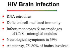
AIDS RELATED INFECTIONS
Acquired immunodeficiency
syndrome (AIDS) results in neurologic
symptoms in approximately 1/3 of patients.
The most commonly reported CNS
complications include opportunistic fungal,
viral and protozoan infections and
lymphoma. Direct infection of the CNS
with human immunodeficiency virus (HIV)
also occurs. More Recently, highly active
antiretroviral treatment (HAART) has
resulted in changes of the neuropathologic abnormalities, with an overall decreased incidence
of opportunistic cerebral infections, while lymphomas and progressive multifocal
leukoencephalopathy remain unchanged.
![]()
Toxoplasmosis
Toxoplasma gondii, a protozoan, is the most common opportunistic infection in AIDS patients, accounting for between 13.4% and 33% of all CNS complications. The characteristic MR appearance is multiple ring-enhancing lesions located at the cortical-medullary junction, but the basal ganglia and white matter are also frequently involved. The amount of peripheral edema is variable. Earlier in the evolution of the abscesses, the nodule may exhibit more homogeneous enhancement with little mass effect or edema. In general, toxo and fungal abscesses evolve more slowly than bacterial ones, but in immunocompromised individuals they can be quite aggressive. Dual infections are common is AIDS patients. In such cases, invariably one of the pathogens is toxoplasma gondii. Toxoplasmosis may also co-exist with lymphoma. Another confounding fact is that the inflammatory reaction to toxoplasmosis may mimic lymphoma on biopsy or CSF cytology.
Cryptococcosis
Cryptococcosis is the most common CNS fungal infection in AIDS, occurring in 8.7-13% of patients. Cryptococcus neoformans has a peculiar propensity to affect individuals
with cell mediated immunity, and it usually produces a meningitis. CSF antibody titers are
not always reliable for diagnosis because the immune response in AIDS patients is so variable.
Imaging studies may be negative or show only mild ventricular dilatation. Since cerebral
atrophy is common in AIDS patients, distinguishing central atrophy from hydrocephalus is
not always easy, and sometimes followup studies or correlation with the clinical picture is
necessary. The cryptococcal organisms may enter the brain via the VR spaces at the base of
the brain. Proliferation of the organisms within the VR spaces produces gelatinous
pseudocysts of variable size to give a mottled appearance on imaging studies. Meningeal
enhancement is not often present unless a chronic inflammation has developed. A chronic
relapsing infection can result in cryptococcal brain abscesses.
![]()
Progressive Multifocal Leukoencephalopathy (PML)
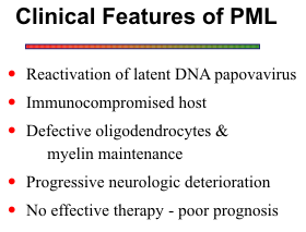
PML is a disorder characterized by widespread foci of demyelination caused by
reactivation of a latent papovavirus. Most cases occur in the setting of an
immunocompromised host secondary to neoplasia, chemotherapy and increasingly, AIDS.
The lesions are initially round or oval, becoming larger and more confluent with time.
Subcortical white matter may be the first
area of involvement with later spread to the
deeper white matter. The pattern is often
asymmetric. The MR appearance reflects
the long T1 and long T2 of the lesions,
typically without mass effect. The high T2
signal is related to both demyelination and
edema. These lesions do not usually
enhance on CT, and experience with MR
shows that most do not enhance with
gadolinium.
![]()
HIV/CMV Encephalitis
Research interests have centered on
a subacute encephalitis involving the white
matter because of its association with HIV infection of the brain and correlation with a
primary AIDS dementia complex. Subacute encephalitis is seen in up to 30% of patients with
AIDS, and cytomegalovirus (CMV) may be the causal agent in some cases. The two viruses
often coexist in brain specimens taken from AIDS patients. In studies of patients with HIV
encephalitis, both CT and MR have been found to be relatively insensitive to the early stages
of involvement, manifested pathologically by widespread microscopic microglial nodules with
multinucleated giant cells. Later, bilateral patchy to confluent lesions in the white matter
develop that are readily visible on MR.
![]()
REFERENCES