DEGENERATIVE AND METABOLIC DISORDERS
DEGENERATIVE DISEASES
Imaging of degenerative disorders with CT has generally been disappointing, and attempts
have been made to apply MR to this area with hopes of demonstrating more specific findings. The
key to the MR imaging of many of these disorders may rest in understanding the normal and
pathologic distribution of iron in the brain. Iron is visualized as areas of hypointensity on T2-weighted
and GRE images caused by local field inhomogeneity and magnetic susceptibility effects. Drayer and
colleagues
![]() noted decreased signal in the globus pallidus, reticular substantia nigra, red nucleus, and
dentate nucleus. These areas correlated closely with sites of preferential accumulation of ferric iron
on Perls' stains in normal brains post mortem. This iron deposition becomes greater with increasing
age, with iron stains first becoming positive at 6 months in the globus pallidum and at 3 to 7 years
in the dentate nucleus. With advanced age (approximately the ninth decade), there may be enough
iron deposition in the putamen to render it as hypointense as the globus pallidus.
noted decreased signal in the globus pallidus, reticular substantia nigra, red nucleus, and
dentate nucleus. These areas correlated closely with sites of preferential accumulation of ferric iron
on Perls' stains in normal brains post mortem. This iron deposition becomes greater with increasing
age, with iron stains first becoming positive at 6 months in the globus pallidum and at 3 to 7 years
in the dentate nucleus. With advanced age (approximately the ninth decade), there may be enough
iron deposition in the putamen to render it as hypointense as the globus pallidus.
Iron may play a role in neurotransmitter metabolism, and several degenerative disorders have
been reported to be associated with increased iron deposition in the brain. This has been described
in Hallervorden-Spatz disease, Huntington's chorea, Parkinson's disease and multisystem atrophy
variants, Alzheimer's disease, and multiple sclerosis.
![]()
Parkinsonism
Parkinson's disease begins most frequently in persons between 40 and 70 years of age. The
characteristic features include slowness of movement, poverty of facial expression, flexed posture,
immobility, and static tremor. Pathologically, there is a loss of pigmented cells in the pars compacta
of the substantia nigra. Parkinson plus syndromes include patients with more severe symptoms and
lesser responses to drug therapy. These syndromes include striatonigral degeneration, Shy-Drager
orthostatic hypotension (multiple system atrophy), olivopontocerebellar atrophy, and progressive
supranuclear palsy.
![]()
Generalized atrophy with prominent sulci and arachnoid spaces is a common finding in these
patients, and the only finding identified with CT in most cases. MR has revealed areas of
hypointensity, which appear to correlate with sites of iron deposition. Findings that have been
described include hypointensity of the putamen, a return to normal signal intensity rather than the
usual low signal intensity of the dorsolateral aspect of the substantia nigra, and narrowing of the band
of relatively increased signal between the hypointense red nucleus and the pars reticulata of the
substantia nigra. This last finding corresponds anatomically with the pars compacta of the substantia
nigra. Varying levels of significance have been reported for these findings in different studies, with
the putaminal hypointensity being least significant and the narrowing of the pars compacta appearing
to be the most significant.
![]()
Initial studies suggested that the putaminal changes were more frequent in Parkinson plus
syndromes, but this has not been found to be the case in a study by Rutledge and associates.
![]() In
progressive supranuclear palsy, atrophy of the midbrain is present. Tissue loss involving the medulla,
pons, brachium pontis, and cerebellum is noted in olivopontocerebellar atrophy.
In
progressive supranuclear palsy, atrophy of the midbrain is present. Tissue loss involving the medulla,
pons, brachium pontis, and cerebellum is noted in olivopontocerebellar atrophy.
Alzheimer's disease
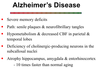
Alzheimer's disease (including senile and presenile forms) is the most common and important
of the degenerative diseases of the brain. Most affected patients are in their fifties or sixties. The
incidence of moderate to severe dementia over the age of 60 has been estimated to be approximately
5 per cent, and 60 per cent of these cases are the result of Alzheimer's disease. Disorders of memory
are first noted, with language disturbances and visuospatial disorientation following. CT reveals
enlargement of the ventricular system and cortical sulci, but this is usually not significantly different
from that in age matched controls. The real utility of CT in Alzheimer's disease lies in the exclusion
of other treatable disorders. MR reveals the same atrophic changes but can better image focal
enlargement of the temporal horns of the lateral ventricles, correlating with hippocampal atrophy .
This atrophy is best seen in the coronal plane. Areas of increased T2 signal were identified in 5 of 12
patients with Alzheimer's disease by Fazekas and associates.
![]() In that particular study, there was no
difference in the frequency of periventricular and deep white matter T2-hyperintense lesions between
controls and patients with Alzheimer's disease. A more extensive smooth halo of periventricular
hyperintensity was noted in 50 per cent of
patients with Alzheimer's disease compared
with controls. Although there are few
specific findings in Alzheimer's disease, the
absence of white matter abnormality,
hydrocephalus, mass lesion, or metabolic
disorder in a demented patient strongly
indicates Alzheimer's or Parkinson's
disease.
In that particular study, there was no
difference in the frequency of periventricular and deep white matter T2-hyperintense lesions between
controls and patients with Alzheimer's disease. A more extensive smooth halo of periventricular
hyperintensity was noted in 50 per cent of
patients with Alzheimer's disease compared
with controls. Although there are few
specific findings in Alzheimer's disease, the
absence of white matter abnormality,
hydrocephalus, mass lesion, or metabolic
disorder in a demented patient strongly
indicates Alzheimer's or Parkinson's
disease.
![]()
On proton spectroscopy, the brains of patients with Alzheimer’s reveal elevated myo-inositol. This finding may not be sufficiently sensitive for diagnosing early Alzheimer’s, but it may be a useful marker for assessing response to new therapies.
Pick’s Disease
Pick's disease or frontotemporal dementia has symptoms largely indistinguishable from those of Alzheimer's disease, although focal disturbances may be more common in Pick's disease. There is striking atrophy of both gray and white matter, typically involving the inferior frontal and temporal lobes.
Creutzfeldt-Jacob Disease
Creutzfeldt-Jakob disease (CJD) is a rapidly progressive degenerative disease caused by a small protein called a prion. Most cases occur sporadically, approximately 10-15% are inherited, or it can be acquired by eating beef containing the infectious prion particle. The clinical picture is one of rapid cognitive decline with psychosis and delirium, and death usually ensues within one year. Diagnosis is made by brain biopsy.
The classic MR findings are symmetric high signal in the basal ganglia on T2 and FLAIR
images and restricted diffusion on diffusion-weighted scans.
![]() Thalamic and cortical involvement can
also occur.
Thalamic and cortical involvement can
also occur.
![]() Later, profound atrophy develops.
Later, profound atrophy develops.
Huntington's Chorea
Huntington's chorea is characterized by a dominant inheritance of dementia and
choreoathetosis. It most often manifests in the fourth and fifth decades. Atrophy of the head of the
caudate nucleus and putamen bilaterally and moderate frontotemporal atrophy produce findings
identifiable with both CT and MR.
![]() The ease of coronal imaging with MR and the greater gray-white matter contrast result in some advantages over CT in diagnosing this condition, but there are
no specific MR signal abnormalities in Huntington's chorea.
The ease of coronal imaging with MR and the greater gray-white matter contrast result in some advantages over CT in diagnosing this condition, but there are
no specific MR signal abnormalities in Huntington's chorea.
METABOLIC DISORDERS
This group includes a long list of diseases that affect the gray and white matter to varying
degrees. This section discusses briefly the major diseases that affect primarily the white matter,
including the classic leukodystrophies. The names and terminologies of these disorders are confusing
because they were derived from the pathologic literature before their metabolic defects were
discovered. As the specific biochemical and enzyme defects are being elucidated, these diseases are
being classified more appropriately. The ones with "cause unknown" are listed under the general
category of primary white matter disorders.
![]()
The classic leukodystrophies include adrenoleukodystrophy, Krabbe's globoid cell, and
metachromatic leukodystrophy, and a few other less well known entities. They have in common a
genetic origin and involve the peripheral nerves as well as the central nervous system. Each is caused
by a specific inherited biochemical defect in the metabolism of myelin proteolipids that results in
abnormal accumulation of a metabolite in brain tissue. Progressive visual failure, mental
deterioration, and spastic paralysis develop early in life, however, variants of these diseases have a
more delayed onset and a less progressive course. The other primary white matter disorders include
Alexander's disease, Canavan disease, Cockayne's syndrome, and Pelizaeus-Merzbacher's disease.
![]()
All of the above white matter diseases are characterized by symmetric massive involvement
of the white matter. MR imaging is very sensitive for detecting the white matter damage, but it is not
very specific. Van der Knaap and his group
![]() developed a computer based pattern recognition
program in attempt to enhance the specificity of image interpretation. Their program uses
information about brain structures involved, lesion characteristics, and special features, such as
calcification, ventricular size, and enhancement with gadolinium.
developed a computer based pattern recognition
program in attempt to enhance the specificity of image interpretation. Their program uses
information about brain structures involved, lesion characteristics, and special features, such as
calcification, ventricular size, and enhancement with gadolinium.
Adrenoleukodystrophy
Adrenoleukodystrophy is a peroxisomal disorder that results in abnormal accumulation of very
long chain fatty acids. Several forms have been described, but x-linked adrenoleukodystrophy is the
classic form that presents in males between the ages of 4 and 8. The neurologic findings of visual and
behavioral problems, intellectual impairment and long tract signs can appear before or after adrenal
gland insufficiency. Adrenoleukodystrophy is both a demyelinating and dysmyelinating disorder.
Initially, it involves predominantly the parietal-occipital lobes and posterior visual pathways, but it
extends forward into the frontal and temporal lobes as the disease progresses. Unlike the focal
plaque-like character of multiple sclerosis, adrenoleukodystrophy tends to be contiguous within fiber
tracts and often is confluent within the larger white matter bundles of the centrum semiovale.
![]() Both
periventricular and subcortical white matter are affected, and in advanced disease the internal capsule,
corpus callosum, corticospinal tracts and other white matter fiber tracts in the brain stem can be
involved.
Both
periventricular and subcortical white matter are affected, and in advanced disease the internal capsule,
corpus callosum, corticospinal tracts and other white matter fiber tracts in the brain stem can be
involved.
The typical MR findings are large, symmetric, hyperintense lesions on T2-weighted images that
are also clearly visible as hypointense areas on T1-weighted scans. The white matter abnormalities
tend to be confluent and of homogeneous signal intensity. Sites of active demyelination along the
advancing edges may be associated with blood-brain barrier disruption and enhance with
paramagnetic contrast agents. Atypical features include frontal lobe involvement, unilateral
involvement, calcifications and mass effect.
![]()
Krabbe's Disease (Globoid Cell Leukodystrophy)
This autosomal recessive disorder presents shortly after birth and progresses rapidly. A
deficiency of the enzyme galactocerebroside beta-galactosidase is the lysosomal defect that results
in profound loss of myelin and destruction of oligodendrocytes. Production and maintenance of
myelin is deficient; the little myelin that is formed is normal morphologically and biochemically.
Characteristic globoid cells with crystalloid cytoplasmic inclusions and extensive reactive gliosis
accompany the deficient myelination histologically. Macrocephaly and marked ventricular dilatation
are other features of Krabbe's disease. MR images reveal bilateral, confluent involvement of the
cerebral and cerebellar white matter. The margins of the lesions may enhance.
![]() ,
,
![]()
Early increased attenuation on CT scans has been noted in the cerebellum, brain stem,
thalamus, caudate, and corona radiata. These areas disclose corresponding hypointensity on T2-weighted scans and normal to hyperintensity on T1-weighted scans, suggesting a some paramagnetic
effect, probably from calcium deposition in those brain structures.
![]()
Metachromatic Leukodystrophy
This is a lysosomal disorder with autosomal recessive inheritance. A deficiency of
arylsulfatase A results in accumulation of sulfatides in the brain and other organ systems. It is
primarily a dysmyelinating disorder, shows no predilection for the parietal-occipital white matter, and
initially spares the arcuate fibers. The cerebellar white matter is commonly affected. Overall, the
distribution is symmetrical, and the lesions of metachromatic leukodystrophy do not enhance. Some
patients exhibit T2 shortening in the thalamus, posterior limb of internal capsule, cerebellum, and
quadrigeminal plate, which has been attributed to a paramagnetic effect from elevated levels of micro-dispersed iron due to dopamine depletion.
![]()
Alexander's Disease
No biochemical marker has been identified in Alexander's disease, so the diagnosis must be
established by clinical criteria and brain biopsy. Astrocytic eosinophilic Rosenthal fibers are the
characteristic histologic findings. It presents early in infancy with psychomotor retardation, and a
progressive downhill course ensues with seizures and spasticity.
![]() It has two distinctive imaging
features, namely macrocephaly and a predilection for the frontal white matter. Contrast enhancement
can be seen during the acute phases of dysmyelination and demyelination. In late stage disease more
global involvement of the white matter is the rule, and brain atrophy also develops.
It has two distinctive imaging
features, namely macrocephaly and a predilection for the frontal white matter. Contrast enhancement
can be seen during the acute phases of dysmyelination and demyelination. In late stage disease more
global involvement of the white matter is the rule, and brain atrophy also develops.
![]()
Canavan’s Disease (Spongiform Leukodystrophy)
Canavan disease is an autosomal recessive disorder, and an enzymatic defect in N-acetylaspartylase has been identified. Vacuoles distributed throughout the white matter gives it a
spongy appearance on histologic examination. Macrocephaly may be the first clinical clue, the MR
scan may show dramatic bilateral white matter abnormality before 1 year of age and before the
neurological deficits become apparent. Rapidly progressive demyelination leads to severe motor and
mental retardation, blindness, and a fatal outcome by 3 years of age. The MR appearance is
nonspecific and similar to the other hereditary disorders.
![]() Proton spectroscopy shows markedly
elevated NAA (N-acetyl aspartate), which is quite specific for Canavan’s disease.
Proton spectroscopy shows markedly
elevated NAA (N-acetyl aspartate), which is quite specific for Canavan’s disease.
Pelizaeus-Merzbacher Disease
No biochemical marker has been discovered in this x-linked recessive disorder. The dominant
feature is profound hypomyelination of the cerebral hemispheres and brain stem secondary to deficient
production of myelin proteins. The presence of no or very little myelin suggests an arrest of
myelination before or shortly after birth. Spared areas of normal myelin in the perivascular regions
produces a characteristic tigroid appearance pathologically that is reflected on MR images as
scattered foci of T2 hyperintensity.
![]() Bilateral symmetric involvement of the white matter and brain
atrophy are nonspecific findings. Hypointensity in basal ganglia and thalamus on T2-weighted scans
suggests pathologic iron storage.
Bilateral symmetric involvement of the white matter and brain
atrophy are nonspecific findings. Hypointensity in basal ganglia and thalamus on T2-weighted scans
suggests pathologic iron storage.
![]()
Cockayne's Syndrome
The hallmark of this disorder is diffuse hypomyelination similar to Pelizaeus-Merzbacher
disease. Sparing of the perivascular myelin is also a feature of this disease. The specific metabolic
defect has not been determined, but the genetic transmission appears to be autosomal recessive. MR
images demonstrate patchy, confluent T2 hyperintensity in the white matter of the cerebral and
cerebellar hemispheres without lobar preference. One distinguishing feature is calcification of the
basal ganglia, dentate nucleus, and periventricular white matter.
![]() ,
,
![]()
Two metabolic disorders, Hurler's disease and Lowe's syndrome, are associated with cystic
changes in the cerebral white matter. Hurler's disease is one of the mucopolysaccharidoses, caused
by an enzymatic defect in the degradation of heparan, dermatan, or keratan sulfate. The cystic
lacunar lesions in the white matter are dilated perineuronal spaces filled with mucopolysaccharide
gargoyle cells.
![]() Lowe's syndrome, one of the aminoacidurias also known as the oculocerebral renal
syndrome, results from defects in the amino acid transport mechanism. In addition to the renal
deficiencies, these patients develop ocular problems and patchy white matter lesions with cystic
components.
Lowe's syndrome, one of the aminoacidurias also known as the oculocerebral renal
syndrome, results from defects in the amino acid transport mechanism. In addition to the renal
deficiencies, these patients develop ocular problems and patchy white matter lesions with cystic
components.
![]()
Leigh’s Disease
Leigh's Disease (subacute necrotizing encephalomyelopathy) is a familial disorder with
autosomal recessive inheritance. Onset is usually in the first year of life in more than half of cases but
occasionally it can present in adulthood. Presenting signs and symptoms range from hypotonia,
seizures and myoclonic jerks in the first year of life to ataxia, dysarthria and nystagmus in the second
year. Death, most often from respiratory failure, usually occurs before 3 years of age. The exact
biochemical defect remains unknown but may involve pyruvate metabolism. Bilaterally symmetric
foci of necrosis and spongiform degeneration are noted pathologically. CT has revealed symmetric
areas of decreased attenuation in the basal ganglia, brain stem and cerebellum. MR has shown these
same lesions as areas of increased T2-signal and has also shown involvement of the tectum,
tegmentum and medullary olive in instances when CT has been negative in these areas.
![]()
Leigh’s disease and other mitochondrial encephalopathies, such as MERRF (mitochondrial
encephalopathy with red ragged fibers) and MELAS (mitochondrial encephalopathy with lactic
acidosis and strokes) show increased brain lactic on proton MR spectroscopy.
![]()
Wilson’s Disease
Wilson’s disease (hepatolenticular degeneration) is an autosomal recessive disorder of copper
metabolism. Onset of symptoms is usually during the second or third decades. The classic syndrome
is dysphagia, slowness and rigidity of movements, dysarthria and tremor. Pathologic changes
primarily involve the lentiform nuclei and range from frank cavitation to softening and atrophy. MR
has demonstrated abnormally increased T2-signal in the putamen and caudate most commonly but
also in the thalamus, dentate nuclei, midbrain and subcortical white matter.
![]()
Hallervorden-Spatz Disease
Hallervorden-Spatz disease is a progressive movement disorder in which there is abnormal
iron deposition in the globus pallidus, reticular zone of the substantia nigra and red nucleus. It is
inherited as an autosomal recessive trait. Onset is in late childhood or early adolescence with both
corticospinal and pyramidal motor findings. MR has revealed decreased T2-signal in the lentiform
nuclei and perilentiform white matter, related to this excess iron deposition. Areas of increased signal
in the periventricular white matter have been noted and these may correlate with disordered
myelination. Disproportionate atrophy of the brainstem and cerebellum is also seen.
![]()
References