SUBARACHNOID HEMORRHAGE
AND INTRACRANIAL ANEURYSMS
John R. Hesselink, MD, FACR
The majority of intracranial aneurysms are congenital or berry aneurysms which occur at the bifurcations of medium-sized arteries at the base of the brain. Arteriosclerotic aneurysms usually have a fusiform shape; traumatic, mycotic and neoplastic aneurysms are much less common and occur in smaller distal vessels.
The incidence of congenital aneurysms in the general population is about 1-2%. If a patient has an unruptured aneurysm noted by CT or angiography, the risk of bleeding is about 1-2% per year. Clinically, a ruptured aneurysm presents as sudden onset of severe headache. In cases of subarachnoid hemorrhages, the most common aneurysms are posterior communicating (38%), anterior communicating (36%), and middle cerebral (21%). These three locations account for 95% of all ruptured aneurysms. The basilar artery accounts for only 2.8% and posterior fossa aneurysms are even less common.

CT of Subarachnoid Hemorrhage
The CT scan is important, first of all, to document the
subarachnoid hemorrhage and to assess the amount of
blood in the cisterns. Detection of subarachnoid blood is
very dependent on how early the scan is obtained. Data in
the literature vary from 60-90%. If the scan is obtained
within four to five days, the detection rate is very high.
Secondly, the CT helps localize the site of the aneurysm.
This can be done by the distribution of blood within the
cisterns and also with dynamic scanning following an IV
bolus of contrast. Thirdly, the CT is important to evaluate
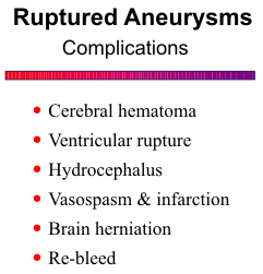
complicating factors such as cerebral hematoma,
ventricular rupture, hydrocephalus, cerebral infarction,
impending uncal herniation and re-bleed.
![]()
Regarding CT patterns of ruptured aneurysm, an anterior communicating aneurysm is suggested by blood in the cisterna lamina terminalis, anterior pericallosal cistern, and interhemispheric fissure. Identification of clot within a cistern makes this sign more specific. There may be extension of blood into the septum pellucidum and lateral ventricle, and hematoma in the inferomedial frontal lobe. Localizing posterior communicating artery aneurysms is more difficult because the blood is usually diffuse within the cisterns. Intracerebral hematoma or ventricular rupture is unusual with posterior communicating aneurysms. Rupture of a middle cerebral aneurysm is characterized by blood in the sylvian fissure and a hematoma in the temporal lobe, which may also rupture into the adjacent temporal horn. Posterior fossa aneurysms often do not have good localizing findings on the CT scan.
It is not uncommon to find a small amount of blood in the ventricles in patients with subarachnoid hemorrhage. That does not necessarily mean that direct ventricular rupture has occurred because subarachnoid blood can enter the ventricular system in a retrograde manner. Ventricular rupture from a bleeding aneurysm is usually more dramatic, often showing a cast of blood or clot in a lateral ventricle. A subarachnoid hemorrhage with blood in the lateral ventricle is usually due to an anterior communicating aneurysm. Middle cerebral aneurysm is another possibility, but that should be associated with a temporal hematoma. Similarly, pericallosal aneurysms can rupture into the ventricle but then there should be hematoma in the corpus callosum as well.

What is the role of a contrast scan in subarachnoid hemorrhage? The combination of clinical and plain scan findings is often fairly conclusive that a subarachnoid hemorrhage has occurred. If emergency arteriography is considered, contrast limitations need to be considered. We obtain the contrast scan if the diagnosis is in doubt, or if the plain scan shows a large intracerebral hematoma that needs emergency evacuation and there is no time for the angiogram. The detection rate of aneurysms with contrast scanning ranges from 40% for posterior communicating to 80% for anterior communicating, middle cerebral and basilar aneurysms. A common problem is that the subarachnoid blood obscures the enhancing aneurysm.
Conventional MR sequences are very insensitive for
detecting subarachnoid hemorrhage. Clots within cisterns
can be detected, but in general, MR is not the procedure of
choice in the work-up of patients with subarachnoid
hemorrhage. Due to the flow void phenomenon, aneurysms
about the circle of Willis can be identified on spin-echo MR images.
![]() With fluid-attenuated inversion
recovery (FLAIR) sequences, the CSF is dark, so that subarachnoid hemorrhage can be seen more
easily. These sequences may be helpful for detecting subarachnoid blood in the posterior fossa where
CT has difficulty and in the sulci over the cerebral convexities.
With fluid-attenuated inversion
recovery (FLAIR) sequences, the CSF is dark, so that subarachnoid hemorrhage can be seen more
easily. These sequences may be helpful for detecting subarachnoid blood in the posterior fossa where
CT has difficulty and in the sulci over the cerebral convexities.
![]()
Giant Aneurysms
Giant aneurysms are a special category defined as any aneurysm larger than 2.5 cm in diameter.
They occasionally bleed but their symptoms are usually related to mass effect. A giant aneurysm
appears on the CT scan as a well-circumscribed mass located at the base of the brain with little or no
surrounding edema. It may have a calcified rim. Thrombus is often found within it, but a patent
lumen can frequently be demonstrated on the contrast scan. The perimeter of the aneurysm may also
enhance.
![]() Giant aneurysms have mixed signal intensity on MR due to a combination of subacute and
chronic hemorrhage and calcification. The patent lumen of the aneurysm may be high or low signal
depending on the rate of flow.
Giant aneurysms have mixed signal intensity on MR due to a combination of subacute and
chronic hemorrhage and calcification. The patent lumen of the aneurysm may be high or low signal
depending on the rate of flow.
![]()
Arteriography
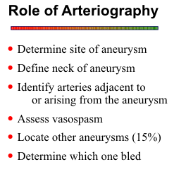
Conventional catheter arteriography still has a vital role in the evaluation of aneurysm and
subarachnoid hemorrhage. The arteriogram is essential, first of all, to determine the site of aneurysm.
CT and clinical findings can suggest the location of the aneurysm, but the arteriogram is still needed
to confirm the specific location of the aneurysm. The information from the clinical and CT findings
should be used to guide the sequence of vessels catheterized during the angiogram. Secondly, the
angiogram must define the neck of the aneurysm so the surgeon can plan his approach for surgical

clipping. Thirdly, it is important to identify arteries
adjacent to or arising from the aneurysm. This
information will also help the surgeon in positioning his
clip, thus avoiding inadvertent clipping of a branch vessel.
Fourthly, assessment of vasospasm is essential for
planning the timing of the operation. Finally, in any
patient with an aneurysm, there is a 15 - 20% chance of
finding multiple aneurysms. For that reason, a three- or
four-vessel angiogram should always be performed in
patients with subarachnoid hemorrhage, even though an
aneurysm might be found on the first vessel studied. In
cases of multiple aneurysms, the angiogram may help
determine which one bled. The angiographic evidence
for the ruptured aneurysm includes the larger aneurysm,
multilobulation and adjacent vasospasm and mass effect.
Again, the CT pattern of subarachnoid blood may also suggest the site of bleeding.
![]()
MR Angiography
MRA can be used to screen for intracranial aneurysms in asymptomatic patients who have a
family history of aneurysms or who have polycystic kidneys, coarctation of the aorta, or collagen
vascular disease, putting them at a higher risk for aneurysms.
![]() 3D time-of-flight (TOF) or phase-contrast (PC) is recommended for screening for aneurysms around the circle of Willis. Relatively
small imaging volumes can be used to keep imaging times short. Aneurysms 5 mm and larger are
detected reliably on good quality studies. Depending on the size of the aneurysm neck and local
hemodynamics, the entire aneurysm lumen may be rendered hyperintense, or an internal flow jet may
be observed similar to the first film of a conventional angiographic sequence.
3D time-of-flight (TOF) or phase-contrast (PC) is recommended for screening for aneurysms around the circle of Willis. Relatively
small imaging volumes can be used to keep imaging times short. Aneurysms 5 mm and larger are
detected reliably on good quality studies. Depending on the size of the aneurysm neck and local
hemodynamics, the entire aneurysm lumen may be rendered hyperintense, or an internal flow jet may
be observed similar to the first film of a conventional angiographic sequence.
![]() The flow jets can be
seen as high or low signal intensity, depending on the flow dynamics and the MRA parameters used.
Potentially slow flow areas, such as the anterior and posterior communicating arteries, may not be
visualized. The resolution of MRA is insufficient to accurately define the aneurysm neck or adjacent
perforating arteries for presurgical planning.
The flow jets can be
seen as high or low signal intensity, depending on the flow dynamics and the MRA parameters used.
Potentially slow flow areas, such as the anterior and posterior communicating arteries, may not be
visualized. The resolution of MRA is insufficient to accurately define the aneurysm neck or adjacent
perforating arteries for presurgical planning.
To avoid errors in diagnosis, all images should be reviewed, including the individual 2D sections
or partitions, the collapse image, and the projection images of the MRA, as well as any available spin-echo scans. Using this approach to screen for aneurysms about the circle of Willis, a sensitivity of
86-95% can be achieved for aneurysms 5 mm or larger,
![]() but the sensitivity decreases to 56% for
smaller aneurysms.
but the sensitivity decreases to 56% for
smaller aneurysms.
![]() Patients must be informed that a normal MRA does not absolutely exclude an
aneurysm. Conventional angiography remains the definitive procedure. For the same reason, MRA
is not recommended in patients with documented subarachnoid hemorrhage.
Patients must be informed that a normal MRA does not absolutely exclude an
aneurysm. Conventional angiography remains the definitive procedure. For the same reason, MRA
is not recommended in patients with documented subarachnoid hemorrhage.
Giant aneurysms are usually readily apparent on spin-echo images. MRA can help identify any residual patent lumen and the parent artery. As mentioned before, intraluminal clot and flow stasis may cause signal loss and obscure the vascular anatomy. Spin saturation is especially a problem for TOF sequences in the larger aneurysms, but spin-phase shifts from swirling blood within the patent lumen also cause signal drop-out and flow-related artifacts in PC images. Also, since the MIP algorithm selects for maximum intensities, an aneurysm that is faintly visible on the collapse image, may not be displayed on the projection images.
CT Angiography
CT angiography is used mostly to define the aneurysm neck and the relationships of adjacent arteries to the aneurysm before therapeutic intervention. Following a bolus injection of 80-100 ml of iodinated contrast at 3-4 ml/min, a helical acquisition is performed through the circle of Willis and any other areas of interest. Images are reconstructed with a 1.25 mm slice thickness at 0.6-1.0 mm increments. In addition to these source images, 3D rendered images and subvolume reformations are helpful to define the aneurysm more accurately.
REFERENCES